Oak Crest Services
Imaging Core
The Imaging Core is a resource for microscopy applications, specialized preparation techniques, immunolabeling, and the expertise to guide and troubleshoot biomedical and environmental research projects to seamlessly integrate imaging data.
Our service supports the local community by providing easy access to high-resolution imaging techniques. We provide contract research services in specimen preparation and imaging, ranging from simple imaging to complete sample preparation, multi-scale correlative imaging and data analysis. We also integrate training in our research and service projects whenever possible and most microscopes are available for independent use to external users.
Published Service Projects
Peng YJ, Geng J, Wu Y, Pinales C, Langen J, Chang YC, Buser C, Chang KT. Minibrain kinase and calcineurin coordinate activity-dependent bulk endocytosis through synaptojanin. J Cell Biol. 2021 Dec 6;220(12):e202011028. doi: 10.1083/jcb.202011028. Epub 2021 Oct 1. PMID: 34596663.
https://doi.org/10.1083/jcb.202011028
Bao X, Zhang Z, Guo Y, Buser C, Kochounian H, Wu N, Li X, He S, Sun B, Ross-Cisneros FN, Sadun AA, Huang L, Zhao M, Fong HKW. Human RGR Gene and Associated Features of Age-Related Macular Degeneration Revealed in Models of Retina-Choriocapillaris Atrophy. Am J Pathol. 2021 May 19:S0002-9440(21)00202-9. doi: 10.1016/j.ajpath.2021.05.003. Epub ahead of print. PMID: 34022179. https://www.sciencedirect.com/science/article/pii/S0002944021002029
Perry S, Goel P, Tran NL, Pinales C, Buser C, Miller DL, Ganetzky B, Dickman D. Developmental arrest of Drosophila larvae elicits presynaptic depression and enables prolonged studies of neurodegeneration. Development. 2020 May 21;147(10):dev186312. doi: 10.1242/dev.186312. PMID: 32345746. https://dev.biologists.org/content/147/10/dev186312
Thornton SM, Samararatne VD, Skeate JG, Buser C, Lühen KP, Taylor JR, Da Silva DM, Kast WM. The Essential Role of anxA2 in Langerhans Cell Birbeck Granules Formation. Cells. 2020; 9(4):974. https://www.mdpi.com/2073-4409/9/4/974
Kelly P, Backes N, Mohler K, Buser C, Kavoor A, Rinehart J, Phillips G, Ibba M. Alanyl-tRNA Synthetase Quality Control Prevents Global Dysregulation of the Escherichia coli Proteome. mBio. 2019 Dec 17;10(6):e02921-19. doi: 10.1128/mBio.02921-19. PMID: 31848288; PMCID: PMC6918089. https://mbio.asm.org/content/10/6/e02921-19
Ding K, Han Y, Seid TW, Buser C, Karigo T, Zhang S, Dickman DK, Anderson DJ. Imaging neuropeptide release at synapses with a genetically engineered reporter. Elife. 2019 Jun 26;8:e46421. doi: 10.7554/eLife.46421. Erratum in: Elife. 2019 Dec 11;8: PMID: 31241464; PMCID: PMC6609332. https://elifesciences.org/articles/46421

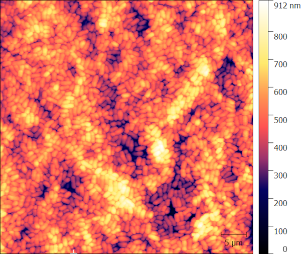
Atomic Force Microscopy (AFM) of a bacterial colony picked off an Agar plate.
Request Services

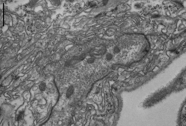
Transmission Electron Microscopy (TEM) of neuromuscular junctions in Drosophila larvae (bar = 500 nm).

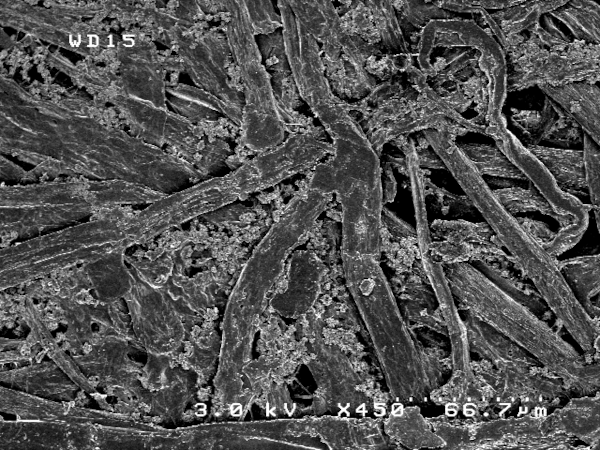
Scanning Electron Microscopy (SEM) of printer paper showing cellulose fibers and additives.
